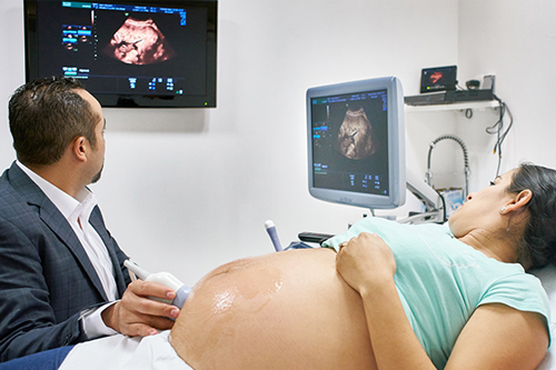
"Fetal 3D viewing" usually refers to the use of 3D ultrasound imaging during pregnancy to get detailed, lifelike pictures of the fetus in the womb. It’s a more advanced version of the traditional 2D ultrasound and is often done during the second or third trimester (around 26–32 weeks) when there’s enough development to see facial features and movement clearly.
2D Ultrasound: Standard black-and-white imaging used to check growth, heartbeat, and anatomy.
3D Ultrasound : Provides a static three-dimensional image of the fetus—great for seeing facial features or body shape.
4D Ultrasound : Like 3D, but with real-time motion (basically a video of the baby moving in 3D).
Medical Reasons : Sometimes used to examine possible abnormalities (e.g. cleft lip or spinal issues).
Keepsake Imaging : Many parents get it for bonding or to keep images/videos as memories.Generally considered safe when done by licensed professionals and within medical guidelines.
Most 3D/4D ultrasounds are non-diagnostic when done at private clinics, meaning they’re not analyzing medical issues—just giving you a keepsake.